Ophthalmologists have long known the connection between genetics and eye care. We have routinely performed “genetic surveys” of nearly every patient we have ever seen, as a standard of care. This may come as a shock to most of my colleagues, especially those who have never had any training in genetics and are not familiar with common DNA terms. Yet, how many of us routinely ask our patients, “Do you have a family history of this illness?” We have accepted the importance of genetic information all along, we just call it by a more conventional term: family history — an important, but unreliable, source of information.
As genetic testing technology has advanced in the last decade, we are now capable of asking a patient about their family history, but then confirming that information objectively by looking at genetic markers that predispose patients to certain diseases. This new capability opens the doors to new approaches to preventative medicine and monitoring that would otherwise not be considered in many patients.
Genetic Testing for the Eye
There have been advancements in genetic markers for eye disease for at least the last 40 years. In the early days, a significant focus was on gene expression and the potential impact on cells, and most of the research breakthroughs focused on the rarest of eye conditions. For example, in 1993 there was excitement around a French team locating the mutation linked to Stargardt’s disease, and by 1998 there was approximately $70 million dollars in grants to the National Eye Institute (NEI) for genetic research into eye diseases.1,2 The Human Genome Project (1990-2003) drove increased awareness and funding, and the eye benefited as well.
Today there is an entire arm of the NEI dedicated to genetics. The identification of rare diseases that have very specific loci was able to advance faster than other diseases. Retinal disorders with targeted loci and their high level of related vision loss especially have received a significant amount of investment and attention; most notably, retinitis pigmentosa (RP) and disorders from mutations in the RPE65 gene that most often afflict children.
The developments in testing, targeting, and therapy resulted in the FDA approval of voretigene neparvovecrzyl (Luxturna, Spark Therapeutics www.sparktx.com) in 2017, the first gene therapy of its type approved anywhere in the world for confirmed biallelic RPE65 mutation-associated retinal dystrophy. While the prevalence of this genetic disease is considerably less than most other eye diseases, the promise that this process of testing and therapy development gives is substantial. There are a number of labs today that offer testing for published eye-related genes, including Arctic Medical Laboratories (www.arcticdx.com ), which offers two types of tests for macular degeneration risk. To find other labs, the Genetic Testing Registry of the National Institutes of Health is available (www.ncbi.nlm.nih.gov/gtr ).
The Cornea Gets its Own Genetic Test
The first commercially available test for corneal dystrophies was made available in Korea by Avellino Labs (www.avellino.com) in 2008. This first test only tested for one TGFBI mutation known to cause Avellino dystrophy (now also known as GCD2). The launch of that test led to more testing in South Korea and Japan and, eventually, more mutations were defined. The “Avellino Test” was modified by Avellino Labs to include five mutations of the TGFBI gene. Today, 42 labs offer corneal disease genetic testing, according to a search on the Genetic Testing Registry. Universities with their own labs, such as Harvard and Baylor, are also able to offer this type of testing.
Initial skepticism in South Korea, and later Japan, over genetic testing for corneal dystrophies subsequently turned into belief, as greater positive results were being found with increased testing. To date, over 770,000 tests have been performed, with over 1,100 positively identified patients. The rate of corneal dystrophies does not make them common diseases by corneal specialists, but the prevalence is significantly greater than the 1:2,000 prevalence ratio that defines a disease as rare, by European Union standards.
In 2019, Avellino Precision Medicine launched the AvaGen test. This test uses next-generation sequencing, also known as NGS, to look for more than 70 mutations of the TGFBI gene for corneal dystrophies, as well as markers across 75 other genes associated with keratoconus (KC) and related diseases. It looks for both corneal conditions at this depth across the identified genetic markers.
Now that we know the technology exists, we need to understand the test’s utility and how we can use it in our practices.
Putting Genetics Into Practice
1 Who are the candidates for corneal genetic testing?
The first candidates to start with are those you have already diagnosed as having the disease. This may seem counterintuitive, but since you know they have progressed already, you can positively identify the genetic markers associated with their progression. KC has a strong link to a positive family history (i.e., genetic link). By identifying the markers that have affected the diagnosed patient, you can now test the family members who may be at risk, especially children and siblings. The fastest progression for KC tends to be between the ages of 13 and 30, and the earlier that these patients can be identified and monitored, the more choices they will have for intervention if the disease shows signs of progressing.
The International Keratoconus Academy (IKA) is doing some excellent work using imaging diagnostics to screen youths in the Chicago area and seeing higher rates of KC than initially expected. These findings are simply based on currently available imaging and corneal curvature measurements. Their testing further proves the theory that the more you test patients, disease detection will occur earlier and more frequently. In the cases of corneal dystrophies and KC, the earlier the detection, the more choices for monitoring, prevention, and potential treatments.
The next patient group is those with elevated mean corneal curvatures and astigmatism rates that demonstrate progression over time, especially concerning in younger patients. There are over 300 million patients globally that could be considered at the pre-clinical stage for KC. Interestingly, more reports are demonstrating that patients can progress well into their 50s and later. Understanding the patient’s genetic predisposition to continued progression of KC could certainly add a dimension to the clinical decision-making process, likened to potentially progressive diseases like glaucoma or AMD. Potential stabilization of this group with cross-linking may also reduce the potential for unpredictable vision changes post-cataract surgery, but that requires a focused study to say for certain.
The final patient groups are those considering corneal refractive surgery with laser vision correction (LVC). As surgeons, we will often encounter “borderline” candidates, and their genetic corneal profile can be an effective additional tool to help determine eligibility for LVC. To date, there is a need for additional prospective studies on these cohorts. Each physician will need to make their own choice to determine which patients they will treat, based on their clinical tools and trained judgement.
An example patient case: A 29-year-old African American male presents for a LASIK evaluation. His Rx has been stable for years, and the patient is too squeamish for contact lens wear. Past medical and family histories are unremarkable:
MRx:
- Right -3.00 +0.50 x164 (20/20)
- Left -3.00 +0.25 x178 (20/20)
- Ultrasound CCT: 505/501 microns
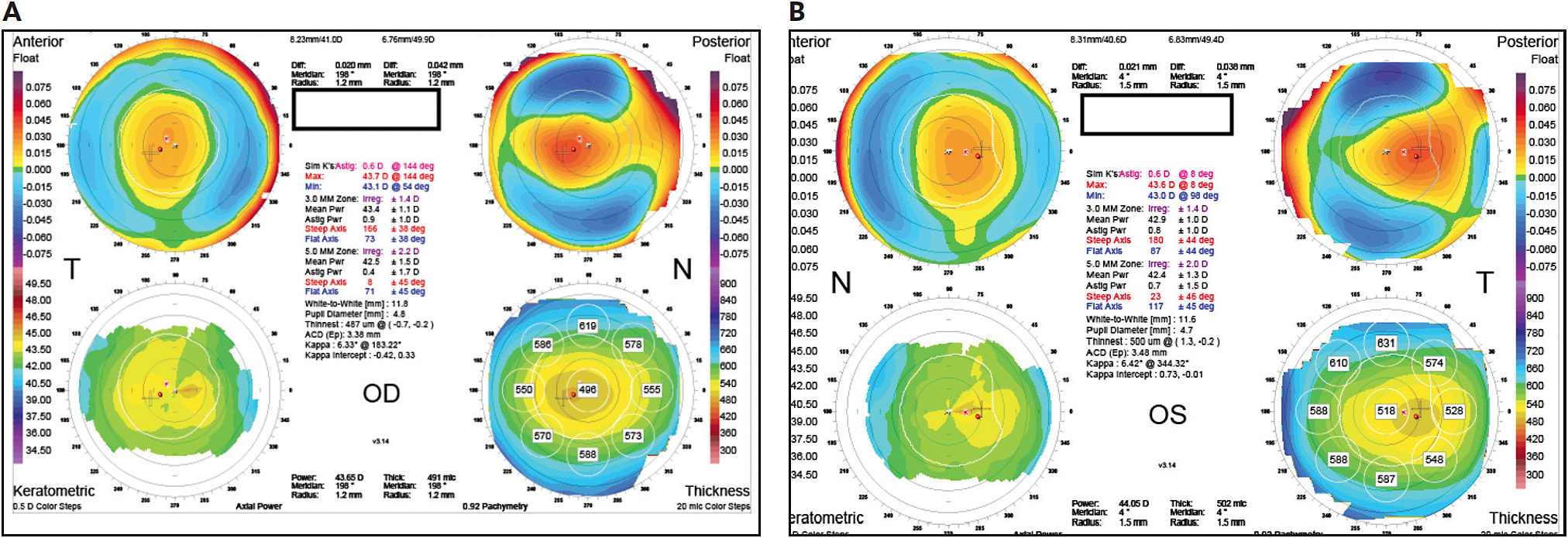
The Orbscan (Bausch + Lomb) images (Figure 1a and Figure 1b) anteriorly demonstrate ~ 0.6 D against-the-rule astigmatism (ATR) in both eyes, with slightly truncated and skewed bowties, left greater than the right eye. The posterior float concerns me a little, as it is trending toward greater elevation in the right eye (0.042 microns), and the left elevation is also slightly more temporal. Personally, anteriorly ATR astigmatism in a young patient puts me on alert, and this in combination with the thinner corneas and the slightly imperfect Orbscan images made this a questionable candidate.

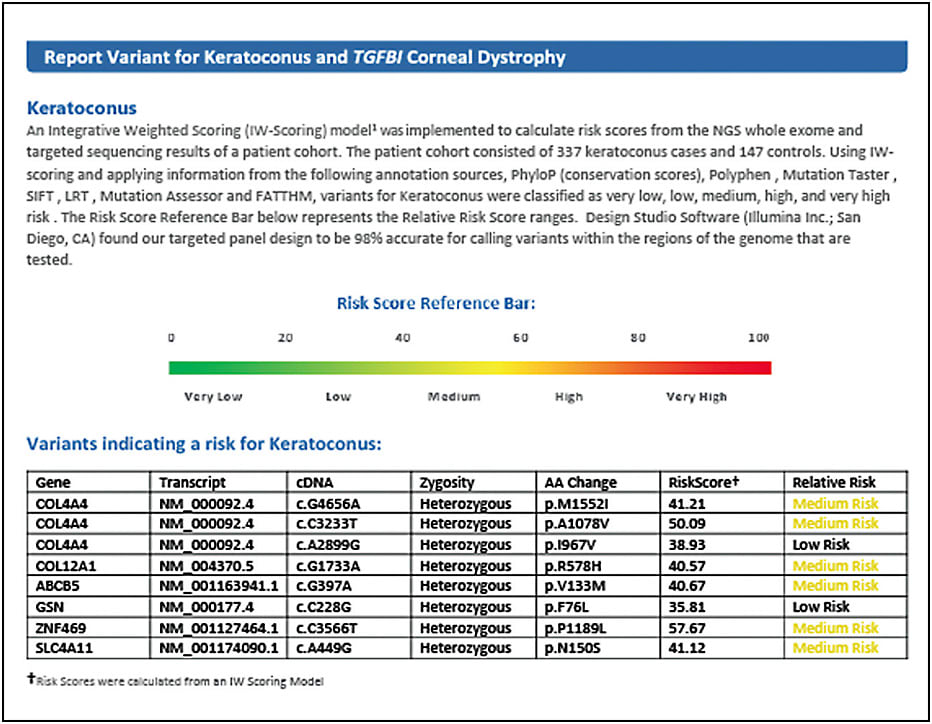
Thus, I recommended a KC genetic screening profile for better insight into this patient’s potential predisposition toward corneal ectasia. Figure 2 demonstrates that this patient has multiple genes that have been present in KC patients. Of particular concern are the “COL” or “collagen-related” genes, with which polymorphism of these particular genes have been seen in up to 4% of KC patients. The cumulative presence of six “medium risk” variants increases this patient’s total risk and, thus, this information “tipped the scale” for me, per se, in my decision-making process toward no laser vision correction at this time.

2 The patients in my practice location are not likely to be affected as much by these diseases. Do I need to use genetic testing?
This is a very common misconception among geography, ethnicity and genetics. With ethnic diversity great and with the very limited genetic knowledge most patients possess, genetic testing is still a very useful tool. The history of GCD2 is a great example: GCD2 was named Avellino corneal dystrophy by a US doctor of European heritage, after he diagnosed the condition in a family from Avellino, Italy who was living near Cincinnati, OH. Also, the test to identify the markers for the disease was developed in South Korea, yet is most prevalent in patients of Chinese descent. The value of genetic testing is the potential for the diagnosis to be a factor in a patient’s treatment options.
3 I don’t have enough time to see patients now, how can I add complex genetic testing to my evaluations and screenings?
This is a major factor for all practices: We are already tight on time to give quality care to each patient, so how do we fit this in? Fortunately, the genetic testing technology has improved so that the sample collection process can be done by a nurse, technician, or trained staff member in the clinic in just a few minutes. The DNA collection is performed by swabbing the buccal mucosa inside of the cheek, and is then deposited into a small vial that preserves the sample. That DNA sample is then shipped to Avellino Precision Medicine, and the reports are accessible to your practice via a secure web portal.
A Valuable Tool
In conclusion, genetic testing is now proving to be an important player for anterior segment surgeons. In particular, KC genetic screening has very unique implications, from preventing unnecessary surgically induced corneal ectasia, to identifying high-risk patients (i.e., family members of KC patients) sooner and screening patients who have high astigmatism earlier and mitigating their disease with collagen cross-linking sooner. The ability to identify gene variants that are related to KC is powerful for clinicians, especially as it relates to potential treatments and follow-up examinations. Even more promising is that this knowledge will ultimately pave the way for creating a truly curative treatment for some of our most challenging corneal disease patients. CP
References:
- Grady D. Gene Discovery May Lead to Test for Devastating Eye Disorder. New York Times. March 4, 1997. https://www.nytimes.com/1997/03/04/science/gene-discovery-may-lead-to-test-for-devastating-eye-disorder.html . Last accessed May 15, 2020.
- Wolkoff L. Researchers track the complex roots of glaucoma in DNA. Ocular Surgery News. 2003; doi https://www.healio.com/ophthalmology/glaucoma/news/print/ocular-surgery-news/%7Bc6acd348-1e62-406d-bfa0-a41c91b8e158%7D/researchers-track-the-complex-roots-of-glaucoma-in-dna .


