Congratulations! You have made the decision to start small incision lenticule extraction, or SMILE, to offer the latest technology in laser vision correction.
SMILE is a relatively new procedure to correct myopia and astigmatism, from -1.00 D to -10.00 D of myopia and 0.75 D to 3.00 D of astigmatism. Its benefits, such as a small side cut, low postoperative total higher-order aberrations, and less risk of postop dry eye disease, have garnered the interest of refractive surgery candidates. Other benefits of SMILE include a “flapless” procedure, increased comfort of the procedure with no laser “odor” and equivalent outcomes compared with LASIK. With current-day practices and laser settings, the recovery is similar to LASIK, with many patients achieving great 1 day postop vision. As a result, surgeons should be armed with action steps to perform this technique successfully. (See “SMILE: An Overview,” below.)
Here, I provide these steps.
Read About SMILE
To learn the “how-to” and nuances of the procedure, seek peer-reviewed journal articles, webinars, videos, and related books. As an example, check out the textbook The Surgeon’s Guide to SMILE: Small Incision Lenticule Extraction, written by Dan Z. Reinstein, MD, Timothy J. Archer, MD, and Glenn I. Carp, MD. Additionally, consider reaching out to colleagues whom you know are well-versed in SMILE for their advice. Using such sources will provide you with an excellent foundation of knowledge.
Get to Know the Laser
Become familiar with data entry, docking, and the laser energy settings. Although you may be an accomplished LASIK surgeon, each femtosecond laser is different, so it is worth taking the time to become familiar with your laser. There can even be differences between and among different VisuMax (Zeiss) lasers. The energy settings for your laser may differ from other lasers in different centers.
SMILE: AN OVERVIEW
Small incision lenticule extraction, or SMILE, involves using a femtosecond laser to create a small side cut, or lenticule, within the cornea. Next, the surgeon uses the laser to make a small, arc-shaped cut on the cornea’s surface to remove the lenticule. This changes the cornea’s shape, correcting the refractive error.
I, personally, review all the data — including refraction, keratometry readings, and pachymetry — that my technician inputs from every patient chart. I have a printed chart note and a tomography for each patient, so it is easy to check for accuracy and/or modify as needed.
The first 50 flaps that you need to complete before starting SMILE (FDA requirement) will give you the opportunity to practice data entry and docking.
Identify Candidates
Ideal candidates for SMILE are at least 22 years old and have not had any changes in their spectacle prescription for at least a year. I find that SMILE works best for my patients who have myopia between -1.75 D to -10.00 D, with less than 2.50 D of astigmatism. Lenticules -1.50 D or less are very thin and, thus, more difficult to dissect. In such cases, I prefer performing LASIK or PRK. Additionally, my LASIK results for patients who have greater than 2.25 D of astigmatism are better than my outcomes with SMILE for this level of astigmatism. I utilize a nomogram for SMILE based on the outcomes of patients with my laser in my center, and add 10% to the spherical component of manifest refraction, and 12% for myopic refractions greater than - 8.00 D. A customized nomogram should be developed for each laser and each center and should be developed based on outcomes.
Patients who have corneal scars should have LASIK rather than SMILE due to concerns about the scars blocking the refractive cut of the laser. Flap creation and lifting is more forgiving with a LASIK flap in the presence of small corneal scars.
For the first 20 cases, I recommend choosing patients who have a prescription of - 4.00 D or greater and a pachymetry of less than 580 μm. This will make dissection easier, as lenticules will not be too thin. Thicker corneas (greater than 580 μm) necessitate dissecting a lenticule in the more anterior stroma, where the collagen fibrils are more densely packed. This makes the dissection more difficult.
Facilitate Lenticule Dissection
In patients whose pachymetry is greater than 580 μm, consider increasing the laser energy by 1 step (5 mJ) across all parameters. The reason: I have found that this makes lenticule dissection easier in thicker corneas. To accomplish this, go to expert settings and adjust the energy levels.
A caveat: Be sure to adjust all categories in the laser settings under “expert settings,” and be sure to re-adjust back to your regular energy settings before your next case. If you have too much of an opaque bubble layer after increasing the energy, decrease the laser energy by 1 step in each category.
Be Aware of Posterior Dissection
If you are having difficulty finding the posterior dissection plane, chances are you may have already dissected posteriorly. To rectify this, perform the anterior rescue maneuver by inserting the Sinskey-type end of your dissector into the lenticule interface. Doing so with a sweeping maneuver near the corners of the entry incision is best; however this can be done at any place in the incision.
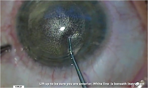
Shining additional light (from a lid speculum light or any other external device) can also help to visualize the lenticule at the incision site. Look for the “white line” of the lenticule — it should not be visible when blocked by a dissecting instrument when the instrument is anterior to the lenticule (i.e., in the anterior plane). (Figure 1). The “white line” of the lenticule should be visible in front of the dissecting instrument when you are in the posterior plane. (Figure 2). Something else to keep in mind: Having your assistant look carefully at the side screen can sometimes provide a clearer view than the direct visualization through the microscope.


Don’t Panic Over Suction Loss
Where I practice, we tape a laminated step-by-step guide to our laser on managing suction loss. Suction loss happens rarely, but usually can be avoided by providing preoperative Diazepam (Pfizer) or a different antianxiolytic agent, along with constant “verbal anesthesia” by reassuring the patient during the laser treatment. I speak to the patient constantly throughout the procedure, letting them know what to expect at each step.
Additionally, I have a nurse or assistant offer to hold the patient’s hand during the procedure for additional comfort. The power of human touch can be very calming.
Examine the Lenticule for Tags
Always assess the lenticule to be sure that you have not left any tags. You can accomplish this by using the slit beam to examine the SMILE treatment zone after lenticule extraction. In addition, examine the lenticule for completeness.
If the treatment zone appears incomplete, use the slit beam and elevate the SMILE treatment zone area with lenticular extraction forceps to look for incomplete tags, as well as fibers, epithelial cells, or meibomian secretions that may have been inadvertently introduced into the interface.
Minimize Rinsing
Reduce rinsing in the interface following lenticule extraction. Doing so will decrease edema and improve early postop visual outcomes.
Have Fun!
Using SMILE is exciting, so enjoy it! It is a great addition to our laser vision correction portfolio. In fact, 90% of my laser vision correction patients receive the SMILE procedure. CP


