Regardless of how many times a surgeon performs keratoplasty, I would argue they still experience the “wow” factor that comes with replacing a diseased cornea with a donor cornea. After all, the procedure enables a patient to regain vision, courtesy of another patient, with the surgeon acting as the vessel. What’s more, the procedure often leads to substantial increases in a patient’s quality of life. All that said, I wonder how many surgeons actually know how this procedure ended up in their hands. If you are curious; you’ll want to read this article. It covers the 3 conceptual periods of keratoplasty:
- Experimentation with xenograft in animal models;
- Success with and the development of penetrating keratoplasty (PK); and
- the addition of selective, layered corneal transplantation.
A caveat: Due to space constraints, it is only possible to paint the broad picture of the development of corneal transplantation. As a result, many of the luminaries in the field, including such individuals as Benjamin Rycroft, Louis Paufique, Tudor Thomas, Max Fine, and Richard Troutman, among many others who made significant contributions, could not be included in this article. Their contributions, however, are no less important in the development of contemporary keratoplasty.
1. Experimentation With Xenograft in Animal Models
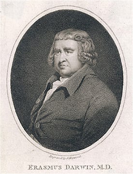
The 18th century was a time of inspiration for the concept of a keratoprosthesis. The first articulated concept of a keratoprosthesis came from Erasmus Darwin, the grandfather of Charles Darwin, in 1798. Specifically, the former Darwin described the use of a glass implant in the cornea of the recipient, though no evidence exists that it was tried.
Similarly, during the French revolution, Guillaume Pellier de Quengsy, in France, described, in detail, a glass keratoprosthesis set in a silver ring, which could replace a scarred cornea. However, once again, no evidence exists that this was attempted.
The period of experimentation and development of both anterior lamellar keratoplasty (ALK), as well as penetrating keratoplasty (PK) took place in the 19th century. The earliest transplants were xenografts. A major proponent of these was Franz Reisinger, from Austria, who coined the term “keratoplasty.”

In addition, Johann Dieffenbach, commonly recognized as the father of strabismus surgery, did extensive experimentation with xenografts in small animals. They were unsuccessful.
Case reports of the procedure also debuted. An example: Richard Sharpe Kissam, of New York, reported using a porcine cornea as a donor for a human who had a scarred cornea, though the keratoplasty attempt was unsuccessful.
In addition, Arthur von Hippel, the chief of surgery at the University of Göttingen’s eye clinic, developed the motorized rotary trephine, revolutionizing the surgical approach to replacement of the cornea. Additionally, he developed ALK as a standard technique.
2. Success With and the Development of PK
It was not until 1906 that Eduard Zirm, in a small outpost in Moravia, reported the first successful PK in a human in the literature, and outlined the principles that underlie that success:
- The use of human donor tissue.
- Strict asepsis.
- Use of the von Hippel rotary trephine.
- Meticulous care of the donor tissue.
- Wound coaptation.
- Use of overlay sutures.
Australian ophthalmologist Anton Elschnig built on the report of Eduard Zirm, performing the first large series of PKs (174), and elaborated on the indications for PK, the preoperative considerations, and the appropriate management of the postoperative course.
Further east, in the Soviet Union,Vladimir Filatov performed a series of 842 PKs between 1922 and 1945. He was the first to use preserved cadaveric donors, and he further developed instruments for trephination and elaborated on a theory of “tissue therapy.”
Ramón Castroviejo, an immigrant from Spain, is credited with largely importing PK to the United States. He developed a practice in New York City and was a passionate advocate of corneal transplantation. Until his death in 1989, Castroviejo was the pre-eminent figure in the moderate era of corneal transplantation and excelled, primarily, as a developer of surgical technique and instrumentation, including corneal trephines, corneal scissors, and forceps to handle cornea tissue.
Practicing across town from Castroviejo was his contemporary, Richard Townley Paton. A graduate of the Wilmer Eye Institute, Paton championed corneal transplantation and established the first eye bank (the Eye-Bank for Sight Restoration) in New York City in 1944.
For the next 75 years, successful PK became a standard of care for patients who had a multiplicity of diseases of the cornea, such as corneal scars from trauma or infection, and opacified corneas from dystrophies or prior surgeries. This procedure was rendered even more successful by the advent of corticosteroids and an enhanced understanding of corneal graft rejection.

3. Corneal Transplantation for Specific Corneal Layers
Interest in the exclusive replacement of the defective layer of the cornea began as early as the 1950s. In fact, José Ignacio Barraquer, in Colombia, and Charles Tillett, in the United States, developed early techniques for posterior lamellar keratoplasty (PLK). However, it was not until 1998 that Gerrit Melles, of the Netherlands, developed a surgical technique for PLK, in which a thin posterior layer of donor cornea with Descemet’s membrane attached was removed from the donor and placed in a trephined pocket of the recipient cornea to provide functional endothelium. This technique was modified and added to by Mark Terry of Portland, Oregon, with improved instrument development to make diseased tissue removal easier. He coined the term “deep lamellar endothelial keratoplasty (DLEK).”
In 2004, Melles introduced the idea of PLK with transplantation of Descemet’s membrane only. This technique was further modified by Francis Price and others to emerge as Descemet’s membrane automated endothelial keratoplasty (DMAEK) in 2006.
The introduction of the “big bubble technique” by Mohammed Anwar has rendered ALK more common in the form of deep anterior lamellar keratoplasty (DALK). In addition to these developments in the early 21st Century, the femtosecond laser has been employed to accomplish “shaped” keratoplasty, which has the theoretical advantage of more rapid healing and better visual outcomes.
From 2010 to the present, there has been a growing interest in cell-based therapy. This includes the introduction of cultured endothelium without a stromal or Descemet carrier into the eyes of patients who have significant endothelial dysfunction from disorders, such as Fuchs’ endothelial dystrophy or pseudophakic corneal decompensation. Shigeru Kinoshita, of Japan, developed this technique and has demonstrated efficacy in a Japanese cohort of patients. At the time of this writing, initial phase 3 studies are being launched in the United States to determine whether cell-based therapy is an effective modality for endothelial replacement.
Descemet’s stripping only (DSO) without transplantation of heterozygous cells was studied and elaborated on by U.S. ophthalmologists Marian Macsai and Kathryn Colby, among others in 2019. In addition, variations on Descemet’s stripping endothelial keratoplasty evolved, including ultra-thin DSAEK, nanothin DSAEK, hemi Descemet’s membrane endothelial keratoplasty, and quarter-DMEK in 2020.

There has been a resurgence in both the interest in and successful performance of prosthokeratoplasty. Newer models of keratoprosthesis designs continue to emerge and may improve patient outcomes based on our understanding of the complications of current devices.
Finally, there is ongoing interest in the development of a biosynthetic cornea, with several centers in collaboration with industry in developing new collagen-based prosthetic approaches.
More “Wow”
This article should provide an even greater “wow” factor when it comes to performing keratoplasty. Consider this: Thanks to keratoplasty’s founding fathers, the present-day corneal surgeon has an expanding repertoire of surgical approaches.
The advent of cell-based therapy, newer types of biosynthetic corneal grafts, novel keratoprostheses, and gene therapy will likely shape the future of surgical approaches to the diseased cornea. CP
NOTE: Dr. Mannis has co-authored “Keratoplasty: a historical perspective,” (Survey of Ophthalmology), among other ophthalmic history articles.


